You Are Here: Home > Recently Diagnosed > Your Pathology Report : Pg. 2

Understanding Your Cancer Pathology Report


Tissue Issues

How is tissue obtained for examination?

In most cases, a doctor needs to do a biopsy or surgery to remove cells or tissues for examination under a microscope.

Biopsies are procedures to collect disease tissue. They are performed in different ways depending on the disease location and other factors.

Some common ways biopsies are done include:

  • Using a needle to withdraw tissue or fluid

  • An endoscope (a thin, lighted tube) to look at areas inside the body and remove cells or tissues

  • Surgery to remove part of the tumor or the entire tumor. If the entire tumor is removed, typically some normal tissue around the tumor is also removed.
 

Tissue removed during a biopsy is sent to a pathology laboratory, where it is sliced into thin sections for viewing under a microscope. This is known as histological (tissue) examination and is usually the best way to tell if cancer is present.

The pathologist may also examine cytologic (cell) material.


Personalized Medicine
 
Recently Diagnosed
 
Cancer 101
 
Survivorship
 
Research
 
Advocacy
 
Inspiration
 
Resources
 
Webinars
 
e-Training
 
Latina Navigator Training


Cytological material is present in:
  • Urine
  • Cerebrospinal fluid (the fluid around the brain and spinal cord)
  • Sputum (mucus from the lungs)
  • Peritoneal (abdominal cavity) fluid
  • Pleural (chest cavity) fluid
  • Cervical/vaginal smears
  • Fluid removed from any biopsy

In a small percentage of cases, an inadequate sample is obtained, which means there is not enough blood or tissue to perform the necessary analysis. In those cases, a second sample must be obtained. It does not mean that an abnormal finding was present, but instead means a laboratory analysis was not performed because the sample size was too small.

How is tissue processed after a biopsy or surgery?

The tissue removed during a biopsy or surgery must be cut into thin sections, placed on slides, and stained with dyes before it can be examined under a microscope.

Two methods are used to make the tissue firm enough to cut into thin sections:

  • Frozen sections
  • Paraffin-embedded (permanent) sections.

All tissue samples are prepared as permanent sections, but sometimes frozen sections are also prepared.

Frozen sections

Are prepared by freezing and slicing the tissue sample. They can be done in about 15 to 20 minutes while the patient is in the operating room. Frozen sections are done when an immediate answer is needed such as to determine whether the tissue is cancerous so as to guide the surgeon during the course of an operation.

Permanent sections

Are prepared by placing the tissue in fixative (usually formalin) to preserve the tissue, processing it through additional solutions, and then placing it in paraffin wax.


After the wax has hardened, the tissue is cut into very thin slices, which are placed on slides and stained.

The process normally takes several days. A permanent section provides the best quality for examination by the pathologist and produces more accurate results than a frozen section.

Image courtesy of the National Cancer Institute (NCI) Visuals Online
 
How long after the tissue sample is taken will the pathology report be ready?

The pathologist sends a pathology report to the doctor within 10 days after the biopsy or surgery is performed. Pathology reports are written in technical medical language. Your doctor should explain the report to you.


Second Opinions

Although some cancers can be easily diagnosed, others cannot. For some types of cancer, a pathology analysis is not an exact science. Depending upon your disease, other pathologists and medical centers might have different opinions on your pathology results or an error by your first Pathologist may have occurred.


A second opinion will require slides and/or paraffin block from the pathologist who examined your sample or from the hospital where your biopsy or surgery was done.

Some cancer centers and other facilities, such as the Armed Forces Institute of Pathology (AFIP), provide second opinions on pathology specimens.


If you want a second opinion, you may wish to contact the facility in advance to determine if this service is available, the cost, and shipping instructions.

Contact information for National Cancer Institute (NCI)-designated cancer centers can be found in the NCI-Designated Cancer Centers database available at:
http://www.cancer.gov/cancertopics/factsheet/NCI/cancer-centers.

Additional information about the AFIP is available on their website at http://www.afip.org.


CISN Tip:

Understanding your pathology report is difficult and not really necessary. But it is important to have a copy of it in your personal records as all next steps are based on this information.

We have included a lot of information here because some people may want to know the details.

And if you want even more technical information about your pathology report, please continue reading the last two questions and answers.


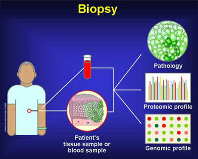
What might the pathology report say about the physical and chemical characteristics of the tissue?

After identifying the tissue as cancerous, the pathologist may perform additional tests to get more information about the tumor that cannot be determined by looking at the tissue with routine stains under a microscope.

The pathology report will include the results of these tests. For example, the pathology report may include information obtained from immunohistochemical stains (IHC). IHC uses antibodies to identify specific antigens on the surface of cancer cells.

IHC can often be used to determine:

  • Where the cancer started,
  • Distinguish among different cancer types: for example, carcinoma, melanoma, and lymphoma
  • Help diagnose and classify leukemias and lymphomas.

The pathology report may also include the results of flow cytometry.

Flow cytometry is a method of measuring properties of cells in a sample, including:

  • The number of cells,
  • The percentage of live cells,
    Cell size and shape
  • Presence of tumor markers on the cell surface. (Tumor markers are substances produced by tumor cells or by other cells in the body in response to cancer or certain noncancerous conditions.)
  • Flow cytometry can be used in the diagnosis, classification, and management of cancers such as acute leukemia, chronic lymphoproliferative disorders, and non-Hodgkin lymphoma.

What information about the genetics of the cells might be included in the pathology report?

Finally, the pathology report may include the results of molecular diagnostic and cytogenetic studies. Such studies investigate the presence or absence of malignant cells, and genetic or molecular abnormalities in specimens.

Cytogenetics uses tissue culture and specialized techniques to provide genetic information about cells, particularly genetic alterations. Some genetic alterations are markers or indicators of a specific cancer.

For example, the Philadelphia chromosome is associated with chronic myelogenous leukemia (CML). Some alterations can provide information about prognosis, which helps the doctor make treatment recommendations.

Some tests that might be performed on a tissue sample include the following:

 
  • Fluorescence in situ hybridization (FISH) determines the positions of particular genes. It can be used to identify chromosomal abnormalities and to map genes.
  • Polymerase chain reaction (PCR) is a method of making many copies of particular DNA sequences of relevance to the diagnosis.
  • Real-time PCR or quantitative PCR is a method of measuring how many copies of a particular DNA sequence are present.
 
Image courtesy of the National Cancer Institute (NCI) Visuals Online
 
  • Reverse-transcriptase polymerase chain reaction (RT-PCR) is a method of making many copies of a specific RNA sequence.
  • Southern blot hybridization detects specific DNA fragments.
  • Western blot hybridization identifies and analyzes proteins or peptides.

  • Microarray testing


1 2
Next Page


Site Design by: Cara M. Caloroso
 
CISN Home Page About Us Services CISN Home Page Contact Site Map CISN Home Page CISN Home Page